Complete cohort - Case 12 of 83
Case 15
Assigned out-of-fold recommendation
Flow diversion alone
Alternative: Coil embolization alone
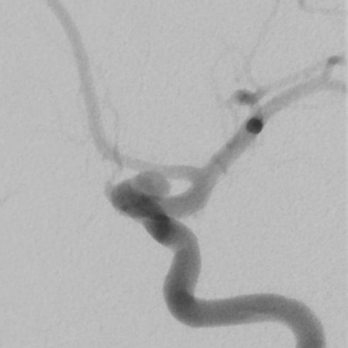
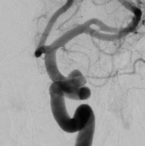
View A is AP; View B is lateral, as confirmed by the data provider.
Case summary
A 67-year-old female presents with an unruptured, incidentally-identified 4.9mm x 4.5mm aneurysm of the left ophthalmic internal carotid artery with a 2.4mm neck.
Image observation
On the supplied views, a laterally projecting saccular outpouching appears at the supraclinoid segment of the internal carotid artery, with the ophthalmic artery origin appearing distinct from the aneurysm fundus.
Model-generated clinical rationale
The assigned recommendation of flow diversion is supported by the aneurysm's location on the ophthalmic segment and the documented separation of the ophthalmic artery origin from the aneurysm fundus, which theoretically allows for parent vessel reconstruction. However, the relatively small aneurysm size and defined neck width also present a viable anatomical target for coil embolization, suggesting the choice between techniques is not strictly dictated by the available morphological data.
Model uncertainty
The OOF classifier showed a relatively clear preference for Flow diversion alone over Coil embolization alone, but its scores are not probabilities of clinical correctness.
Clinical uncertainty / missing factor
The most significant missing decision factor is the specific clinical risk profile regarding the trade-off between the need for dual antiplatelet therapy with flow diversion versus the potential for coil compaction or recurrence.
Explicit evidence used
4.9mm x 4.5mm aneurysm with 2.4mm neck | Ophthalmic artery origin is separate from the aneurysm fundus