Complete cohort - Case 45 of 83
Case 54
Assigned out-of-fold recommendation
Coil embolization alone
Alternative: Stent-assisted coil embolization
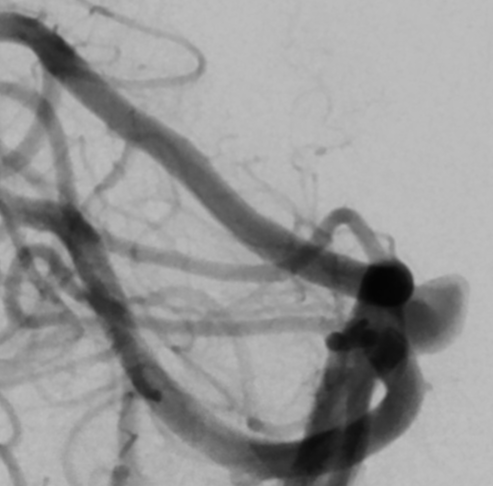
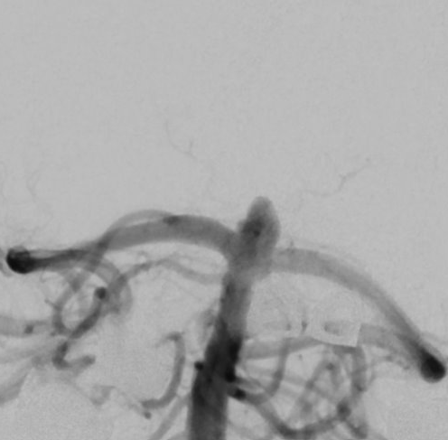
View A is AP; View B is lateral, as confirmed by the data provider.
Case summary
An 80-year-old male presents with an incidental 4.6mm x 4.3mm x 5.6mm superiorly and anteriorly projecting saccular aneurysm at the basilar artery apex.
Image observation
On the supplied views, a saccular outpouching appears at the basilar apex with contrast filling the adjacent posterior cerebral arteries.
Model-generated clinical rationale
The assigned recommendation of coil embolization alone is supported by the aneurysm's saccular morphology and the documented normal caliber of the basilar trunk and posterior cerebral arteries. However, the recommendation is limited by the lack of explicit documentation regarding the aneurysm neck width or the presence of incorporated branch vessels, which are critical factors for determining the feasibility of simple coiling.
Model uncertainty
The OOF score distribution was diffuse: Coil embolization alone ranked first, but Stent-assisted coil embolization remained a material alternative.
Clinical uncertainty / missing factor
The specific neck-to-dome ratio and the precise relationship of the aneurysm neck to the origins of the posterior cerebral arteries are not explicitly quantified in the narrative.
Explicit evidence used
4.6mm x 4.3mm x 5.6mm superiorly and anteriorly projecting saccular aneurysm at the basilar apex | Normal caliber of basilar trunk and posterior cerebral arteries