Complete cohort - Case 3 of 83
Case 6
Assigned out-of-fold recommendation
Coil embolization alone
Alternative: Balloon-assisted coil embolization
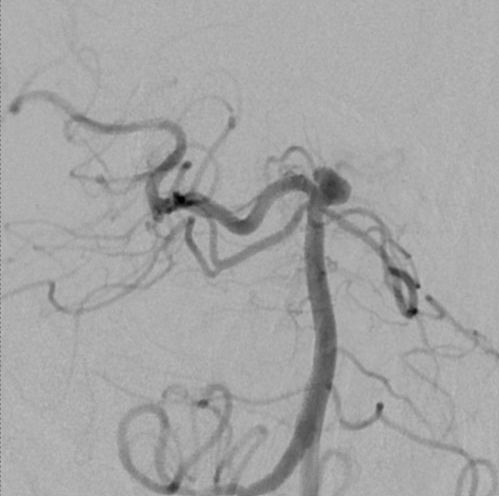
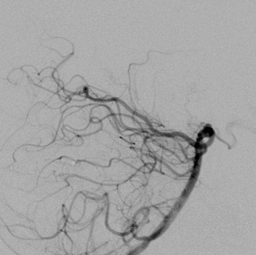
View A is AP; View B is lateral, as confirmed by the data provider.
Case summary
A 55-year-old man with subarachnoid hemorrhage presents with a 5.5mm x 4mm x 3mm irregular, bilobed basilar tip aneurysm and a fetal left posterior communicating artery.
Image observation
On the supplied views, the aneurysm appears as a distinct contrast-filled outpouching at the basilar terminus with adjacent branching vessels visible.
Model-generated clinical rationale
The assigned recommendation of coil embolization alone is supported by the aneurysm's small dimensions and the documented irregular, bilobed morphology, which may facilitate coil retention. However, the complex shape and location at the basilar tip often necessitate adjunctive balloon assistance to maintain coil stability and protect adjacent perforators.
Model uncertainty
The OOF classifier scores for Coil embolization alone and Balloon-assisted coil embolization were close, so the assigned recommendation should be treated as uncertain.
Clinical uncertainty / missing factor
The specific neck width and the precise relationship of the aneurysm to the perforating vessels are not explicitly quantified, limiting the certainty of coil stability without balloon support.
Explicit evidence used
5.5mm x 4mm x 3mm irregular basilar tip aneurysm | Bilobed in appearance