Complete cohort - Case 61 of 83
Case 76
Assigned out-of-fold recommendation
Stent-assisted coil embolization
Alternative: Balloon-assisted coil embolization
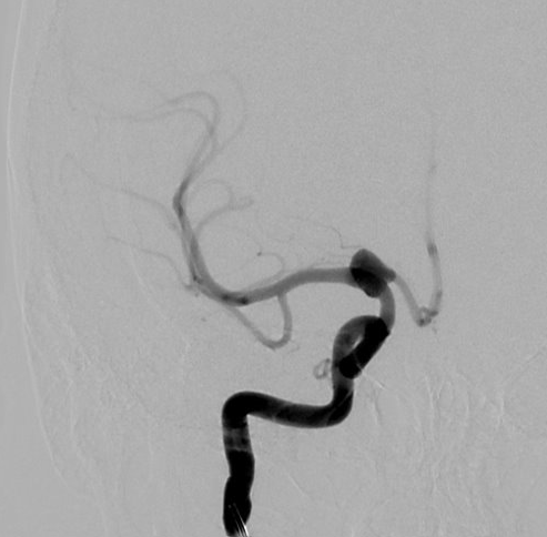
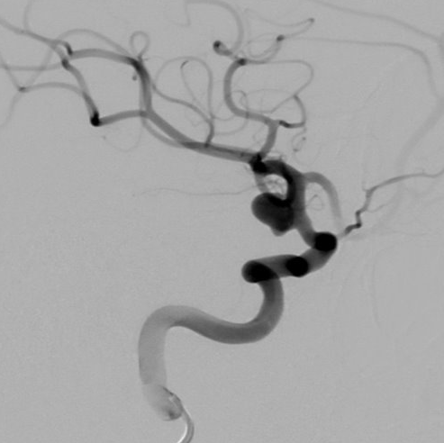
View A is AP; View B is lateral, as confirmed by the data provider.
Case summary
A 69-year-old female presented with an unruptured 6mm x 8mm x 5.38mm right internal carotid artery aneurysm with a 4.2mm neck.
Image observation
On the supplied views, a saccular outpouching appears to arise from the communicating segment of the internal carotid artery, with the parent vessel and distal branches opacifying normally.
Model-generated clinical rationale
The assigned recommendation of stent-assisted coil embolization is supported by the documented 4.2mm neck width, which suggests a wide-necked morphology that may require adjunctive scaffolding for coil stability. However, the rationale is limited by the explicit finding that the posterior communicating artery is not visualized, preventing confirmation of branch incorporation or collateral dependence.
Model uncertainty
The OOF score distribution was diffuse: Stent-assisted coil embolization ranked first, but Balloon-assisted coil embolization remained a material alternative.
Clinical uncertainty / missing factor
The most significant missing decision factor is the visualization of the posterior communicating artery, which is necessary to determine if the aneurysm neck incorporates a branch vessel that would contraindicate stent placement.
Explicit evidence used
6mm x 8mm x 5.38mm aneurysm with 4.2mm neck | Posterior communicating arteries are not visualized