Complete cohort - Case 63 of 83
Case 78
Assigned out-of-fold recommendation
Coil embolization alone
Alternative: Balloon-assisted coil embolization
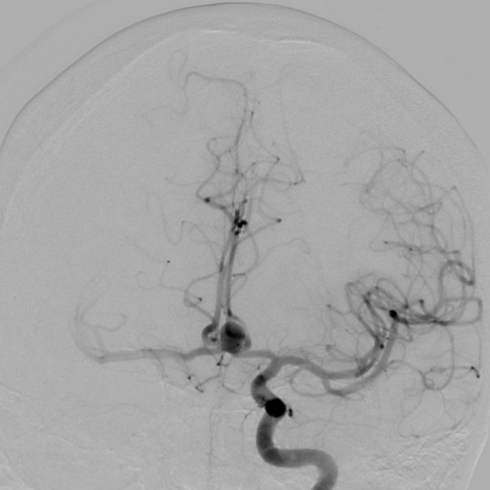
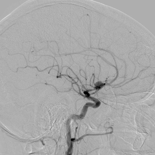
View A is AP; View B is lateral, as confirmed by the data provider.
Case summary
A 69-year-old female presented with subarachnoid hemorrhage and a 1.9mm-necked aneurysm at the anterior communicating artery.
Image observation
On the supplied views, the aneurysm appears as a distinct contrast-filled outpouching at the anterior communicating artery junction.
Model-generated clinical rationale
The documented 1.9mm neck width suggests a geometry that may be amenable to coil embolization alone. However, the narrative notes complex morphology and poor visualization of the neck, which limits confidence in this approach without further anatomical definition.
Model uncertainty
The OOF classifier showed a relatively clear preference for Coil embolization alone over Balloon-assisted coil embolization, but its scores are not probabilities of clinical correctness.
Clinical uncertainty / missing factor
The most significant uncertainty is the precise neck-to-dome ratio and the exact relationship of the A2 segment origins to the aneurysm neck, which are critical for determining coil stability.
Explicit evidence used
1.9mm neck width | Complex morphology and poor visualization of the aneurysm neck