Complete cohort - Case 5 of 83
Case 8
Assigned out-of-fold recommendation
Flow diversion alone
Alternative: Flow diversion with coil embolization
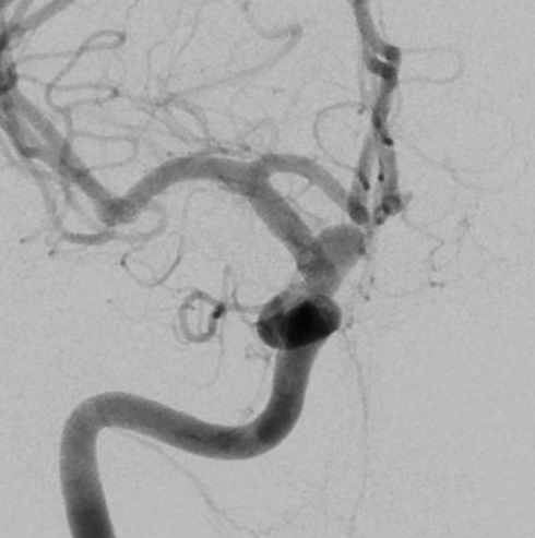
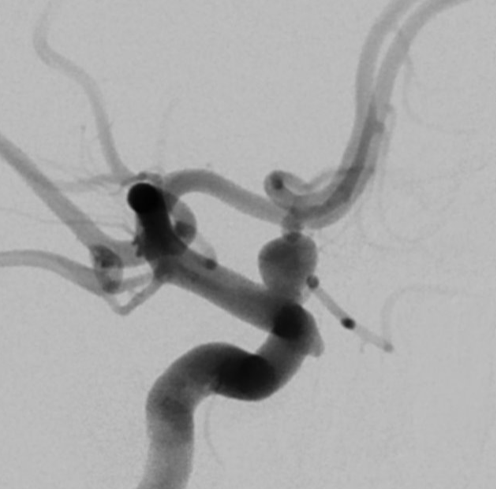
View A is AP; View B is lateral, as confirmed by the data provider.
Case summary
A 44-year-old female presented with a subarachnoid hemorrhage and an incidentally found 5.0mm x 4.9mm right ophthalmic segment aneurysm with a 3.3mm neck.
Image observation
On the supplied views, a saccular outpouching appears at the ophthalmic segment of the internal carotid artery, with the ophthalmic artery arising from the aneurysm base.
Model-generated clinical rationale
The assigned recommendation of flow diversion alone is supported by the aneurysm's location and the presence of a branch vessel arising from the base, which may complicate coil placement. However, the relatively wide neck width of 3.3mm limits the certainty of this approach, as it may require adjunctive coiling to prevent device protrusion or ensure adequate neck coverage.
Model uncertainty
The OOF score distribution was diffuse: Flow diversion alone ranked first, but Flow diversion with coil embolization remained a material alternative.
Clinical uncertainty / missing factor
The most important missing decision factor is the specific clinical status of the subarachnoid hemorrhage, which determines the safety of antiplatelet therapy required for flow diversion.
Explicit evidence used
5.0mm x 4.9mm aneurysm with 3.3mm neck | Ophthalmic artery arises from the base of the aneurysm