Complete cohort - Case 6 of 83
Case 9
Assigned out-of-fold recommendation
Coil embolization alone
Alternative: Stent-assisted coil embolization
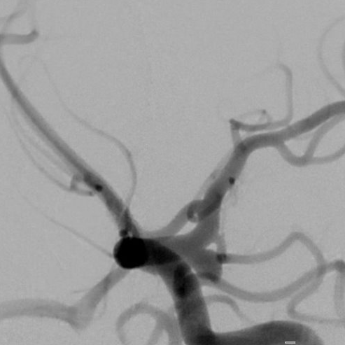
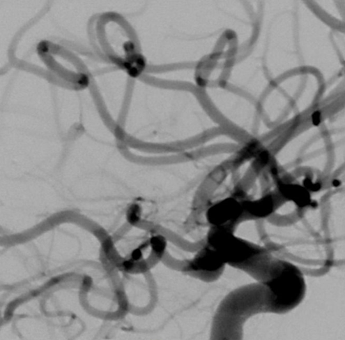
View A is AP; View B is lateral, as confirmed by the data provider.
Case summary
A 46-year-old patient presents with subarachnoid hemorrhage and a 6.9mm x 5.5mm x 7.8mm aneurysm with a 4.8mm neck arising from the left internal carotid artery communicating segment.
Image observation
On the supplied views, a saccular outpouching appears to project posteriorly from the internal carotid artery, with adjacent branch vessels visible in the working projections.
Model-generated clinical rationale
The assigned recommendation of coil embolization alone is supported by the documented aneurysm dimensions and the specific measurement of a 4.8mm neck. However, the relatively wide neck width and the complex morphology noted in the narrative may limit the stability of coils without adjunctive support, making the recommendation potentially questionable.
Model uncertainty
The OOF score distribution was diffuse: Coil embolization alone ranked first, but Stent-assisted coil embolization remained a material alternative.
Clinical uncertainty / missing factor
The definitive visualization of the anterior and posterior communicating arteries is missing, as they were not visualized on the diagnostic angiogram, which limits the assessment of branch incorporation.
Explicit evidence used
6.9mm x 5.5mm x 7.8mm aneurysm with 4.8mm neck | projecting posteriorly from the communicating segment of the left ICA