Complete cohort - Case 7 of 83
Case 10
Assigned out-of-fold recommendation
Coil embolization alone
Alternative: Balloon-assisted coil embolization
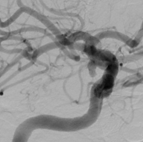
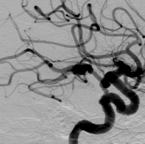
View A is AP; View B is lateral, as confirmed by the data provider.
Case summary
A 68-year-old female with Hunt and Hess 3 subarachnoid hemorrhage presents with a 4.8 x 2.4 mm right PCOM aneurysm featuring a 3.5 mm neck and a large PCOM artery arising from the aneurysm neck.
Image observation
On the supplied views, the aneurysm appears as an elongated, irregular filling defect projecting posteriorly and inferiorly from the right internal carotid artery, with the PCOM artery arising from the aneurysm neck.
Model-generated clinical rationale
The assigned recommendation of coil embolization alone is supported by the aneurysm's oblong morphology and the documented large caliber of the PCOM artery, which may facilitate catheter access. However, the wide 3.5 mm neck and the incorporation of the PCOM origin from the neck present significant challenges for coil stability without adjunctive balloon or stent support.
Model uncertainty
The OOF classifier showed a relatively clear preference for Coil embolization alone over Balloon-assisted coil embolization, but its scores are not probabilities of clinical correctness.
Clinical uncertainty / missing factor
The most critical missing decision factor is the precise 3D relationship of the PCOM origin to the aneurysm neck, which determines the feasibility of preserving the branch without adjunctive devices.
Explicit evidence used
4.8 x 2.4 mm aneurysm with 3.5 mm neck | Large right PCOM arising from the aneurysm neck