Complete cohort - Case 66 of 83
Case 81
Assigned out-of-fold recommendation
Coil embolization alone
Alternative: Balloon-assisted coil embolization
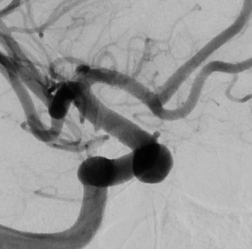
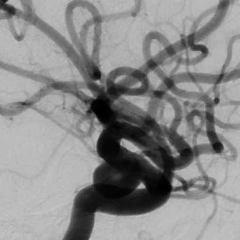
View A is AP; View B is lateral, as confirmed by the data provider.
Case summary
A 37-year-old female patient presents with a 7 mm x 7 mm x 8 mm right supraclinoid internal carotid artery aneurysm projecting medially and dorsally.
Image observation
On the supplied views, the aneurysm appears as a distinct, rounded contrast-filled outpouching arising from the supraclinoid internal carotid artery.
Model-generated clinical rationale
The assigned recommendation of coil embolization alone is supported by the documented absence of a posterior communicating artery and the normal caliber of the anterior choroidal artery, suggesting a lack of critical branch incorporation. However, the specific neck width and dome-to-neck ratio are not explicitly quantified in the narrative, which limits the ability to confirm the feasibility of simple coiling without adjunctive devices.
Model uncertainty
The OOF classifier showed a relatively clear preference for Coil embolization alone over Balloon-assisted coil embolization, but its scores are not probabilities of clinical correctness.
Clinical uncertainty / missing factor
The most significant missing decision factor is the precise aneurysm neck width and dome-to-neck ratio, which are critical for determining the stability of coils without balloon assistance.
Explicit evidence used
7 mm x 7 mm x 8 mm supraclinoid aneurysm | No visualized posterior communicating artery