Complete cohort - Case 73 of 83
Case 88
Assigned out-of-fold recommendation
Coil embolization alone
Alternative: Stent-assisted coil embolization
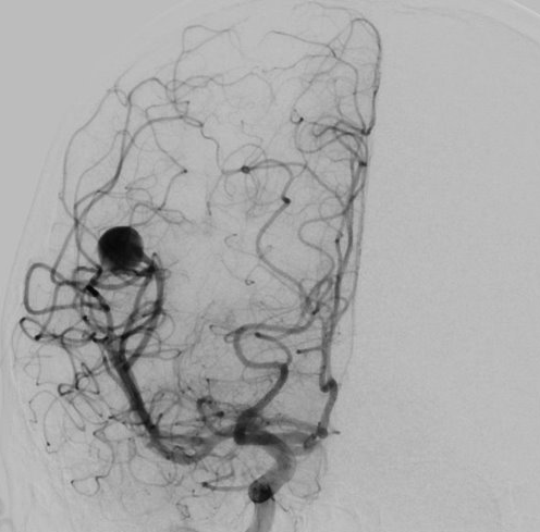
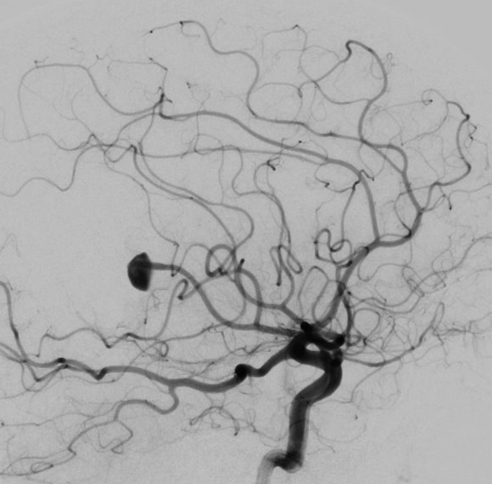
View A is AP; View B is lateral, as confirmed by the data provider.
Case summary
A 70-year-old female presents with an incidental, unruptured 10.7mm x 9.6mm x 8mm aneurysm of the right MCA angular (M3) branch.
Image observation
On the supplied views, a well-circumscribed aneurysm appears at the distal MCA distribution with a visible neck connecting to the parent vessel.
Model-generated clinical rationale
The assigned recommendation of coil embolization alone is supported by the aneurysm's well-circumscribed morphology and distal M3 location, which may allow for stable coil packing without adjunctive devices. However, the large sac dimensions and the presence of significant distal flow delay and avascular regions suggest that the aneurysm neck width and potential branch incorporation remain critical, yet undocumented, factors that could necessitate stent assistance.
Model uncertainty
The OOF score distribution was diffuse: Coil embolization alone ranked first, but Stent-assisted coil embolization remained a material alternative.
Clinical uncertainty / missing factor
The most significant missing decision factor is the precise aneurysm neck width and the exact relationship of the neck to the angular branch origin, which determines the feasibility of coil retention without stenting.
Explicit evidence used
10.7mm x 9.6mm x 8mm well circumscribed aneurysm | Right MCA angular M3 branch location