Complete cohort - Case 18 of 83
Case 22
Assigned out-of-fold recommendation
Coil embolization alone
Alternative: Balloon-assisted coil embolization
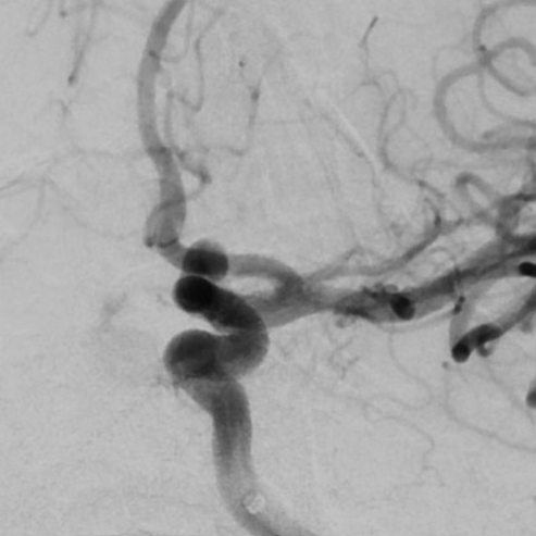
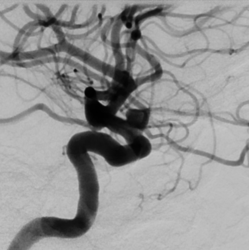
View A is AP; View B is lateral, as confirmed by the data provider.
Case summary
The patient presents with an unruptured 3.5 mm x 3.7 mm x 4 mm left ophthalmic saccular aneurysm with a 3 mm wide neck, from which the ophthalmic artery originates.
Image observation
On the supplied views, a saccular outpouching appears at the ophthalmic segment of the internal carotid artery with a visible branch vessel arising from the neck region.
Model-generated clinical rationale
The assigned recommendation of coil embolization alone is supported by the aneurysm's small dimensions and saccular morphology. However, the documented 3 mm neck width and the origin of the ophthalmic artery from the neck present significant technical challenges for simple coiling, as these features typically necessitate balloon assistance to maintain coil stability and protect the branch vessel.
Model uncertainty
The OOF score distribution was diffuse: Coil embolization alone ranked first, but Balloon-assisted coil embolization remained a material alternative.
Clinical uncertainty / missing factor
The most critical missing decision factor is the specific assessment of the ophthalmic artery's incorporation into the aneurysm neck, which determines the necessity of balloon remodeling.
Explicit evidence used
3.5 mm x 3.7 mm x 4 mm left ophthalmic saccular aneurysm | 3 mm wide neck with ophthalmic artery origin