Complete cohort - Case 21 of 83
Case 25
Assigned out-of-fold recommendation
Coil embolization alone
Alternative: Stent-assisted coil embolization
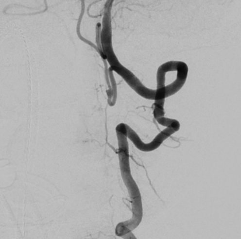
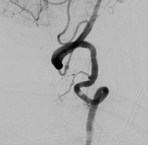
View A is AP; View B is lateral, as confirmed by the data provider.
Case summary
A 79-year-old woman presented with Hunt and Hess 4, Fisher 3 subarachnoid hemorrhage caused by a 2.0 x 2.7 mm fusiform dissecting aneurysm on the proximal cisternal segment of the left PICA.
Image observation
On the supplied views, the left PICA appears tortuous with a focal fusiform dilation on the proximal cisternal segment, while the vertebral artery origin appears extremely tortuous.
Model-generated clinical rationale
The assigned recommendation of coil embolization alone is supported by the small dimensions of the fusiform segment, which may allow for packing without adjunctive devices. However, the dissecting nature of the lesion and the extreme tortuosity of the vertebral artery origin may complicate catheter stability and device delivery. The rationale is limited by the lack of specific documentation regarding the aneurysm neck width or the feasibility of maintaining a stable microcatheter position.
Model uncertainty
The OOF classifier showed a relatively clear preference for Coil embolization alone over Stent-assisted coil embolization, but its scores are not probabilities of clinical correctness.
Clinical uncertainty / missing factor
The most critical missing decision factor is the precise neck morphology and the feasibility of maintaining catheter stability given the extreme vertebral artery tortuosity.
Explicit evidence used
2.0 x 2.7 mm fusiform segment on left PICA | Extremely tortuous vertebral artery origin