Complete cohort - Case 23 of 83
Case 27
Assigned out-of-fold recommendation
Flow diversion with coil embolization
Alternative: Flow diversion alone
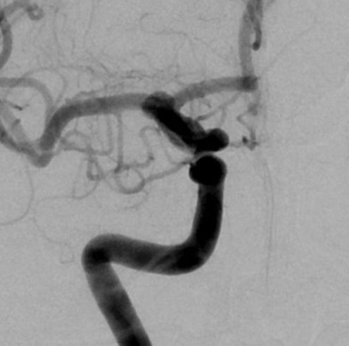
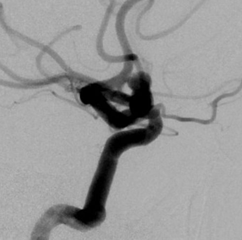
View A is AP; View B is lateral, as confirmed by the data provider.
Case summary
A 41-year-old male presents with a 6 mm x 5 mm x 12 mm partially thrombosed, windsock-shaped unruptured right ophthalmic aneurysm with a 1.2 mm neck.
Image observation
On the supplied views, a large fusiform dilation appears to involve the ophthalmic segment of the internal carotid artery, with the vessel lumen appearing patent.
Model-generated clinical rationale
The assigned recommendation of flow diversion with coil embolization addresses the wide-necked, windsock morphology and partial thrombosis described in the narrative. However, the documented 1.2 mm neck is relatively narrow, which typically favors flow diversion alone; the addition of coils may be intended to manage the thrombus burden or specific geometry, though this is not explicitly confirmed by the images.
Model uncertainty
The OOF classifier scores for Flow diversion with coil embolization and Flow diversion alone were close, so the assigned recommendation should be treated as uncertain.
Clinical uncertainty / missing factor
The decision to combine coils with flow diversion is uncertain given the narrow neck width, and the specific impact of the partial thrombosis on device selection is not fully defined.
Explicit evidence used
6 mm x 5 mm x 12 mm windsock shaped aneurysm | 1.2 mm neck | partially thrombosed