Complete cohort - Case 43 of 83
Case 52
Assigned out-of-fold recommendation
Coil embolization alone
Alternative: Stent-assisted coil embolization
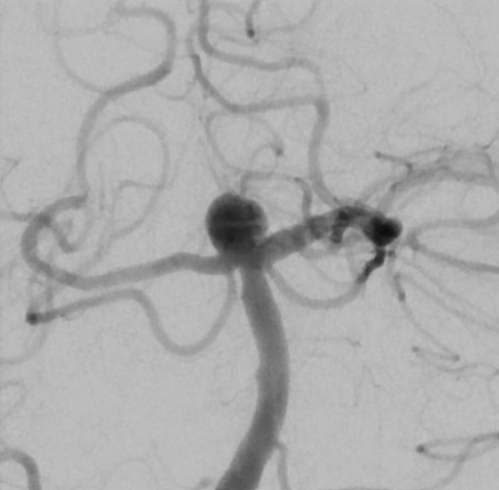
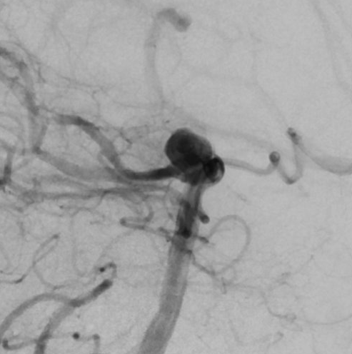
View A is AP; View B is lateral, as confirmed by the data provider.
Case summary
A 60-year-old female presents with a recently discovered unruptured right P1/basilar apex aneurysm measuring 5.3 x 5.6 x 6.5 mm with a 3.6 mm neck.
Image observation
On the supplied views, a saccular aneurysm appears at the basilar apex, projecting superiorly and favoring the right P1 segment.
Model-generated clinical rationale
The assigned recommendation of coil embolization alone is supported by the documented 3.6 mm neck width, which may allow for primary coil packing without adjunctive devices. However, the aneurysm's location at the basilar apex and its dimensions suggest a complex geometry that could challenge coil stability. The recommendation is limited by the lack of explicit documentation regarding the dome-to-neck ratio or specific morphological features that would definitively rule out the need for stent assistance.
Model uncertainty
The OOF score distribution was diffuse: Coil embolization alone ranked first, but Stent-assisted coil embolization remained a material alternative.
Clinical uncertainty / missing factor
The most significant missing decision factor is the precise dome-to-neck ratio and detailed morphological assessment required to confirm the feasibility of primary coiling versus stent assistance.
Explicit evidence used
Unruptured right P1/basilar apex aneurysm | Aneurysm measures 5.3 x 5.6 x 6.5 mm with a 3.6 mm wide neck