Complete cohort - Case 55 of 83
Case 66
Assigned out-of-fold recommendation
Coil embolization alone
Alternative: Flow diversion with coil embolization
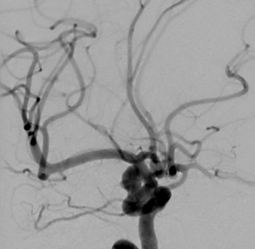
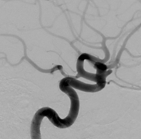
View A is AP; View B is lateral, as confirmed by the data provider.
Case summary
A 56-year-old female presents with an incidental 8.0 mm x 6.4 mm right ICA ophthalmic segment aneurysm with a 4.1 mm neck.
Image observation
On the supplied views, a saccular outpouching appears to project superiorly from the internal carotid artery, with the parent vessel and distal branches appearing patent.
Model-generated clinical rationale
The assigned recommendation of coil embolization alone is supported by the documented 4.1 mm neck width, which may allow for primary coiling. However, the relatively wide neck-to-dome ratio and the aneurysm's location at the ophthalmic segment limit the certainty of this approach, as the risk of coil prolapse or ophthalmic artery compromise remains a concern.
Model uncertainty
The OOF classifier scores for Coil embolization alone and Flow diversion with coil embolization were close, so the assigned recommendation should be treated as uncertain.
Clinical uncertainty / missing factor
The most significant missing decision factor is the precise morphological assessment of the neck and the exact spatial relationship to the ophthalmic artery origin, which are critical for determining the safety of primary coiling versus flow diversion.
Explicit evidence used
8.0 mm x 6.4 mm aneurysm with 4.1 mm neck | Right ICA ophthalmic segment location